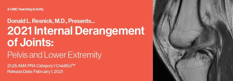
Internal Derangement of Joints 2021: Pelvis and Lower Extremity (CME VIDEOS)
$9
Internal Derangement of Joints 2021: Pelvis and Lower Extremity (CME VIDEOS)
Include: 32 videos 1 pdf, size: 6.08 GB
This continuing education activity provides an update of current information on MR Imaging in the assessment of musculoskeletal disorders. Essential anatomy, physiology and pathology are emphasized that explain imaging findings in disorders of the pelvis, hip, knee, ankle and foot. MR imaging findings in the assessment of common problems in lower extremity joints are compared to those derived from other imaging methods.
Target Audience
The target audience is practicing radiologists, orthopedic surgeons, rheumatologists, podiatrists, sports medicine physicians and other physicians interested in musculoskeletal disorders.
Educational Objectives
At the completion of this CME activity, subscribers should be able to:
– Assess MR images in patients with internal derangements of peripheral joints.
– Articulate the anatomic features fundamental to accurate interpretation of the MR imaging findings in these disorders.
– Formulate a reasonable differential diagnostic list and be able to identify the most likely diagnosis.
– Comprehend the pathogenesis and imaging findings associated with common and important disorders that affect the pelvis and lower extremities.
Topics/Speakers:
ARTICULAR DISORDERS OF SYNOVIUM-LINED JOINTS: Joint Morphology and General Abnormalities, Specific Inflammatory and Degenerative Disorders, Tumors and Tumor-Like Disorders
Donald L. Resnick, M.D.
FOCUS SESSION: Osteonecrosis Versus Insufficiency Fractures: Emphasis on the Hip and Knee
Donald L. Resnick, M.D.
Chondral, Osteochondral and Subchondral Injuries: Anatomy, Pathophysiology, Terminology and MR Imaging
Donald L. Resnick, M.D.
Cartilage Imaging: Routine and Advanced Methods
Christine B. Chung, M.D.
Stress-Related Abnormalities of the Skeleton with Emphasis on the Pelvis and Lower Extremity
Mini N. Pathria, M.D.
FOCUS SESSION: Muscle Disorders: Anatomy Pathophysiology and General Abnormalities
Mini N. Pathria, M.D.
Muscles and Tendons About the Pelvis and Hip: Anatomy, Strains and Tears
Mini N. Pathria, M.D.
Labral Abnormalities and External and Internal Femoroacetabular Impingement
Donald L. Resnick, M.D.
FOCUS SESSION: Important Entrapment Neuropathies of the Pelvis and Lower Extremity
Evelyne A. Fliszar, M.D.
Meniscus: Structure, Function and Patterns of Failure
Donald L. Resnick, M.D.
Discoid Menisci and Other Anomalies
Donald L. Resnick, M.D.
FOCUS SESSION: Bone Marrow: Normal and Abnormal with Emphasis on MRI
Evelyne A. Fliszar, M.D.
Anatomy, Biomechanics and Footprints of Injury
Donald L. Resnick, M.D.
Anterior Cruciate Ligament
Brady Huang, M.D.
Posterior Cruciate Ligament
Brady Huang, M.D.
0.25 Hrs $22.50
Medial Supporting Structures of the Knee
Donald L. Resnick, M.D.
Lateral Supporting Structures of the Knee
Brady Huang, M.D.
Postoperative Ligaments with Emphasis on the Anterior Cruciate Ligament
Brady Huang, M.D.
Patellofemoral Maltracking and Patellar Instability/Dislocation
Lucas Hiller, M.D., M.S.E.
Quadriceps/Patellar Tendons, Fat Pads, Bursae and Plicae
Mini N. Pathria, M.D.
Popliteal Fossa
Mini N. Pathria, M.D.
FOCUS SESSION: MRI/Arthroscopy Correlation
Eric Y. Chang, M.D.
Osteomyelitis, Septic Arthritis and Soft Tissue Infection with Emphasis on the Diabetic Foot
Karen C. Chen, M.D.
0.5 Hrs
Fractures/Dislocations of the Ankle and Foot: Role of CT Scanning
Tudor Hughes, M.D., FRCR
Tumors and Tumor-Like Lesions of the Ankle and Foot
Edward Smitaman, M.D.
Tendons: Normal Anatomy
Donald L. Resnick, M.D.
Adult Acquired Flatfoot Deformity
Mini N. Pathria, M.D.
Tendons: Tendinosis, Tenosynovitis, Tendon Tears and Other Tendon Abnormalities
Donald L. Resnick, M.D.
0.5 Hrs
Rapid Fired Case Review Session
Tarsal Coalition, Osteochondritis Dissecans of the Talus, Metatarsalgia, Plantar Aponeurosis
Edward Smitaman, M.D., Karen C. Chen, M.D., Christine B. Chung, M.D. Karen C. Chen, M.D.
Ligaments: Normal Anatomy
Donald L. Resnick, M.D.
Ligaments: Patterns of Injury
Donald L. Resnick, M.D.
CME Release Date 1/31/2021
Topics:
1 Articular disorders of Synovium-lined joints.mp4
2 FOCUS SESSION Osteonecrosis Versus Insufficiency Fractures.mp4
3 Chondral, Osteochondral and Subchondral Injuries.mp4
4 SAM Session Cartilage Imaging Routine and Advanced Methods.mp4
5 Stress-Related Abnormalities of the Skeleton with Emphasis on the Pelvis and Lower Extremity.mp4
6 FOCUS SESSION Muscle Disorders Anatomy Pathophysiology and General Abnormalities.mp4
7 Muscles and Tendons About the Pelvis and Hip Anatomy, Strains and Tears.mp4
8 Labral Abnormalities and External and Internal Femoroacetabular Impingement.mp4
9 FOCUS SESSION Important Entrapment Neuropathies of the Pelvis and Lower Extremity.mp4
10 Meniscus Structure, Function and Patterns of Failure.mp4
11 Discoid Menisci and Other Anomalies.mp4
12 FOCUS SESSION Bone Marrow Normal and Abnormal with Emphasis on MRI.mp4
12.1 – Old lecture MRI of Bone Marrow Disorders.mp4
13 Knee Anatomy, Biomechanics and Footprints of Injury.mp4
14 Anterior Cruciate Ligament.mp4
15 Posterior Cruciate Ligament.mp4
16 Medial Supporting Structures of the Knee.mp4
17 Lateral Supporting Structures of the Knee.mp4
18 Postoperative Ligaments with Emphasis on the Anterior Cruciate Ligament.mp4
19 Patellofemoral Maltracking and Patellar Instability Dislocation.mp4
20 Quadriceps Patellar Tendons, Fat Pads, Bursae and Plicae.mp4
21 Popliteal Fossa.mp4
22 FOCUS SESSION Knee MRI Arthroscopy Correlation.mp4
23 Osteomyelitis, Septic Arthritis and Soft Tissue Infection with Emphasis on the Diabetic Foot.mp4
24 Fractures Dislocations of the Ankle and Foot Role of CT Scanning.mp4
25 Tumors and Tumor-Like Lesions of the Ankle and Foot.mp4
26 Tendons of Ankle and Foot Normal Anatomy.mp4
27 Adult Acquired Flatfoot Deformity.mp4
28 Tendons Tendinosis, Tenosynovitis, Tendon Tears and Other Tendon Abnormalities.mp4
29 How about Rapid Fired Case Review Session.mp4
30 Ligaments of Ankle and Foot Normal Anatomy.mp4
31 Ligaments of Ankle and Foot Patterns of Injury.mp4
Brochure for lower extremity and pelvis.pdf
Related products